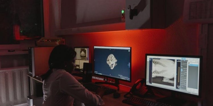
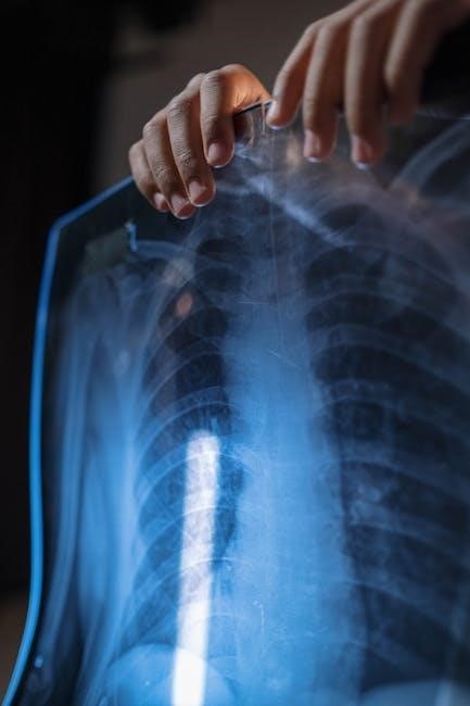
Digital radiography technique charts, often available as a PDF, are essential guides. They detail kVp, mAs, time, and SID for optimal imaging of various body parts.
These resources, like those found on ControlTheDose.com, offer templates for film/screen and digital imaging, aiding radiographers in consistent, quality examinations.
What are Digital Radiography Technique Charts?
Digital radiography technique charts are systematic collections of recommended imaging parameters, frequently distributed as PDF documents, designed to produce diagnostic-quality radiographs. Charts specify settings like kilovoltage peak (kVp), milliampere-seconds (mAs), exposure time, and source-image distance (SID) tailored to specific anatomical regions and patient sizes.
These charts aren’t rigid rules, but rather starting points, acknowledging variations in equipment and patient factors. Resources like ControlTheDose.com provide downloadable PDF versions, including options for both high-frequency and single-phase systems, alongside film/screen and digital modalities. They ensure standardized protocols, promoting image consistency and minimizing unnecessary radiation exposure.
Importance of Standardized Technique Charts
Standardized digital radiography technique charts, often accessed as a convenient PDF, are crucial for maintaining consistent image quality across all examinations. Utilizing these charts minimizes variability between radiographers, leading to more reliable diagnoses. They also play a vital role in dose optimization, ensuring patients receive the lowest possible radiation exposure for a diagnostic image.
Consistent application of these charts, available from resources like ControlTheDose.com, supports quality control and facilitates accurate comparisons of images over time. A well-maintained PDF chart serves as a cornerstone of a robust radiography program, enhancing both patient safety and diagnostic accuracy.
Evolution of Technique Charts: From Film/Screen to Digital
Historically, radiography technique charts were designed for film/screen systems, focusing on optimizing density and contrast. The transition to digital radiography necessitated significant adaptations, as digital detectors exhibit a wider dynamic range and linear response. Consequently, technique charts evolved, shifting emphasis from density control to exposure indicator management.
Modern digital technique charts, frequently available as a PDF, now prioritize achieving optimal signal-to-noise ratio and minimizing patient dose. Resources like ControlTheDose.com provide both legacy and digital-specific charts, reflecting this evolution in imaging technology and practice.
Key Parameters in Digital Radiography Technique Charts
Digital radiography technique charts (often PDFs) detail crucial settings: kVp, mAs, time, and SID. These parameters ensure image quality and dose optimization.
Kilovoltage Peak (kVp) Selection
Kilovoltage peak (kVp) selection, detailed in digital radiography technique charts (often PDF format), profoundly impacts image contrast and penetration. Higher kVp values increase penetration, suitable for thicker body parts like the thorax or pelvis.
Conversely, lower kVp values enhance contrast, ideal for visualizing subtle bone structures in extremities or the skull. Technique charts provide starting points, but radiographers must adjust based on patient size and anatomy.
Proper kVp selection minimizes patient dose while maintaining diagnostic image quality, a core principle highlighted in resources like ControlTheDose.com’s PDF guides.
Milliampere-Seconds (mAs) Determination
Milliampere-seconds (mAs), crucial for image density, is meticulously outlined in digital radiography technique charts, frequently available as PDF downloads. mAs controls the quantity of x-ray photons produced; higher mAs yields darker images, while lower mAs results in lighter images.
Charts offer baseline mAs values, but adjustments are vital based on patient size, body habitus, and the specific anatomy being imaged.
Optimizing mAs is key to balancing image quality and minimizing patient radiation dose, a focus of resources like ControlTheDose.com’s comprehensive PDF technique guides;
Time and Source-Image Distance (SID) Considerations
Digital radiography technique charts, often accessible as a convenient PDF, detail the interplay between exposure time and Source-Image Distance (SID). Altering either impacts image quality and patient dose.
Increasing SID reduces magnification and geometric unsharpness, requiring a longer exposure time to maintain image density. Conversely, decreasing SID necessitates a shorter time.
Charts provide standardized SID values, typically 100cm, but adjustments are sometimes necessary. Understanding this relationship, as outlined in PDF resources like those from ControlTheDose.com, is vital for optimal imaging.
Patient Weight and Body Habitus Impact
Digital radiography technique charts, frequently found as downloadable PDFs, acknowledge the significant influence of patient weight and body habitus on radiation exposure.
Larger patients require increased mAs to penetrate tissues adequately, ensuring sufficient image receptor exposure. Body habitus—the patient’s build—also plays a role; denser builds necessitate higher techniques.
Some charts incorporate weight-based adjustments, while others offer general guidelines. Hoerner, Grizzard, and Moroz’s research highlights methods for determining technique based on weight and height, optimizing dose and image quality, as detailed in available PDF resources.
Anatomy-Specific Technique Charts
Digital radiography technique charts, often in PDF format, provide tailored settings for each body region—skull, thorax, pelvis, and extremities—for optimal imaging.
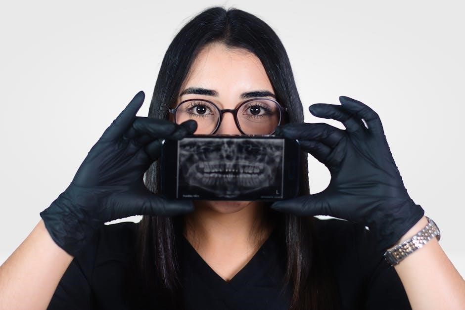
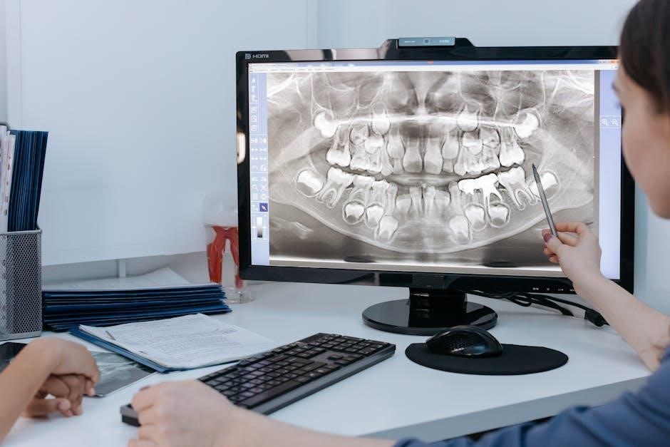
Skull and Facial Bones Radiography Techniques
Digital radiography technique charts, frequently accessible as a PDF download, offer specific guidance for skull and facial bone imaging. These charts detail recommended kVp and mAs settings, varying based on projection—PA, lateral, or Waters view—and anatomical region, like the sinuses.
For example, a standard lateral skull may utilize 70-80 kVp and 3-5 mAs, while facial bone imaging often requires lower kVp (60-70) for enhanced contrast. Proper collimation is crucial to minimize scatter radiation. Charts also consider patient size; larger patients may necessitate increased mAs. Utilizing these PDF resources ensures consistent, high-quality images while adhering to ALARA principles.
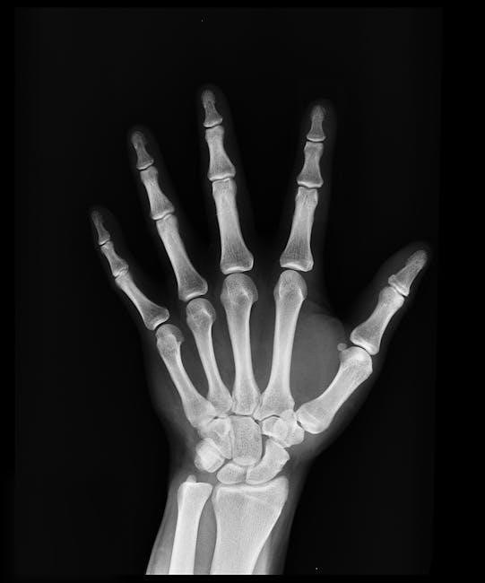
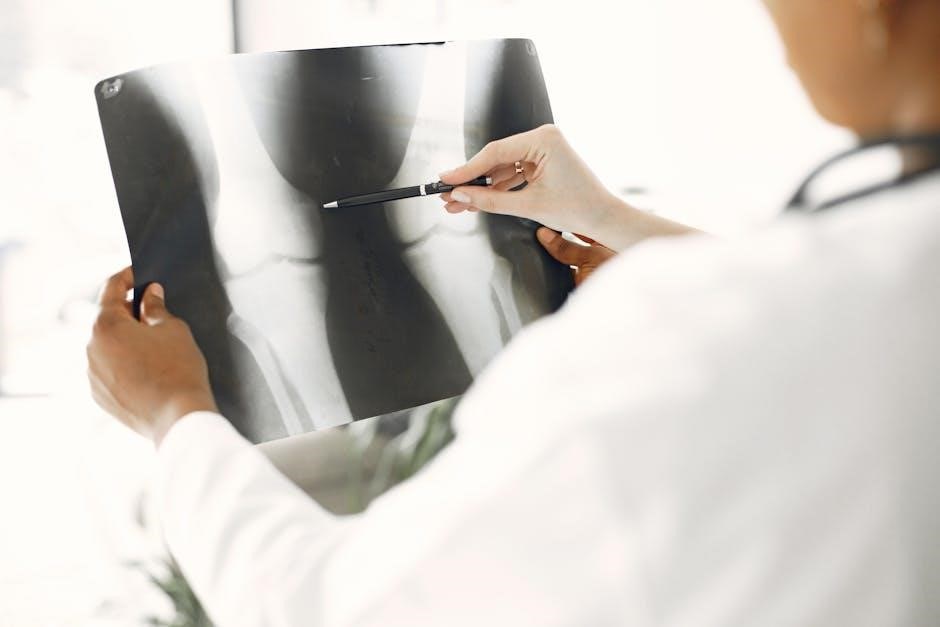
Shoulder Girdle and Extremity Techniques
Digital radiography technique charts, often found as downloadable PDFs, provide essential parameters for imaging the shoulder girdle and extremities. These charts specify kVp (typically 65-80 kVp) and mAs settings, adjusted for body part and patient size.
For instance, shoulder AP projections might use 70 kVp and 2-3 mAs, while hand/wrist imaging often employs lower doses. Charts detail appropriate SID (Source-Image Distance) and collimation techniques. Consideration of patient habitus is vital; larger limbs require increased mAs. These PDF guides promote standardized protocols, optimizing image quality and minimizing radiation exposure.
Thorax (Chest) Radiography Techniques
Digital radiography technique charts, readily available as PDF downloads, are crucial for chest imaging. Standard PA and lateral projections typically utilize 120-140 kVp and adjusted mAs values based on patient size and body habitus.
Portable chest radiographs often require technique adjustments due to increased SID. Charts emphasize collimation to minimize scatter radiation and optimize image quality. AEC (Automatic Exposure Control) usage is detailed, alongside considerations for lung fields and cardiac silhouette visualization. These PDF resources ensure consistent protocols, balancing diagnostic accuracy with ALARA principles – As Low As Reasonably Achievable.
Pelvis and Lower Extremity Techniques
Digital radiography technique charts, often found as convenient PDFs, provide guidelines for pelvic and lower extremity imaging. AP pelvis projections generally employ 100-120 kVp, with mAs adjusted for patient build. Femur and tibia/fibula studies utilize similar kVp ranges, varying mAs based on projection and anatomy.
Ankle and foot imaging requires lower techniques, around 60-80 kVp. Charts detail SID considerations and collimation practices to reduce scatter. These PDF resources emphasize proper positioning and technique selection for optimal visualization of bony structures and joints, ensuring diagnostic image quality.
Hand, Wrist, and Finger Radiography
Digital radiography technique charts, readily available as PDF downloads, are crucial for hand, wrist, and finger imaging. Typically, these examinations utilize lower kVp settings, ranging from 50-70 kVp, to enhance soft tissue contrast. mAs values are adjusted based on patient size and the specific projection—PA, lateral, or oblique.
Finger imaging demands even lower exposure factors. Charts emphasize meticulous collimation to minimize dose and artifact. Proper positioning, detailed in these PDF guides, is vital for accurate assessment of fractures and joint spaces, ensuring diagnostic quality in these small anatomical structures.
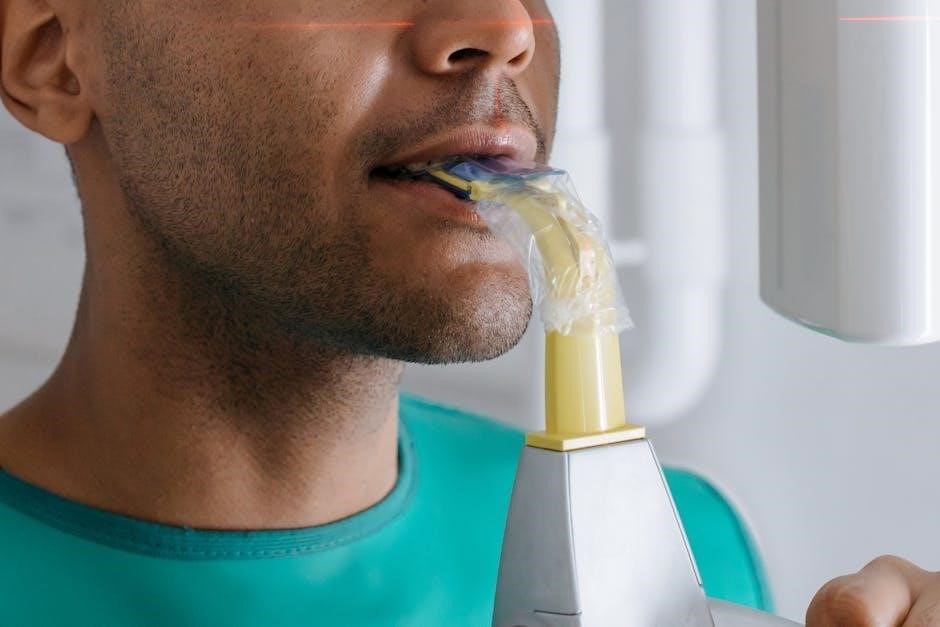
Digital Radiography Systems and Technique Adaptation
Digital systems, like CR and DR, necessitate technique adjustments. PDF technique charts guide these adaptations, considering factors like detector type and AEC utilization for optimal imaging.
Computed Radiography (CR) Technique Considerations
Computed radiography (CR) technique charts, frequently found as downloadable PDF documents, are vital for establishing appropriate exposure parameters. CR systems generally require slightly higher mAs settings compared to direct digital radiography due to the indirect conversion process.
Radiographers must carefully consult these charts, considering patient size and anatomy, to minimize repeat exposures and maintain image quality. Utilizing a PDF technique chart ensures consistency and adherence to established protocols. Proper collimation and beam limitation, as outlined in these resources, are crucial for reducing patient dose and improving image contrast in CR examinations.
Understanding CR system limitations and adapting techniques accordingly, guided by a reliable PDF chart, is paramount for optimal diagnostic results.
Direct Radiography (DR) Technique Considerations
Direct radiography (DR) technique charts, often accessible as a convenient PDF, leverage the system’s direct conversion capabilities for efficient imaging. DR systems typically require lower mAs settings than CR, due to their higher detective quantum efficiency.
Radiographers should utilize PDF technique charts as a foundation, adjusting parameters based on patient habitus and anatomical region. These charts emphasize the importance of precise kVp selection for optimal contrast and detail.
DR’s wider dynamic range allows for greater latitude in exposure, but adherence to standardized charts ensures consistent image quality and minimizes unnecessary radiation dose, as detailed in downloadable PDF resources.
Impact of Automatic Exposure Control (AEC)
Automatic Exposure Control (AEC) significantly influences digital radiography technique, and technique charts, often found as PDF downloads, must account for its use. AEC systems automatically adjust mAs to achieve a predetermined image receptor exposure.
While simplifying exposure selection, radiographers must understand AEC limitations and potential errors. PDF technique charts often provide guidance on AEC chamber selection and factors affecting its performance, like patient size and positioning.
Proper AEC utilization, guided by technique charts, optimizes image quality and minimizes patient dose, ensuring adherence to ALARA principles. Regular quality control checks of AEC are crucial for reliable performance.
Dose Reduction Strategies in Digital Radiography
Digital radiography technique charts (PDF format) promote ALARA principles through collimation, optimal kVp/mAs, and image processing, minimizing patient exposure.
ALARA Principle and Digital Radiography
Digital radiography technique charts, frequently accessed as a convenient PDF, are instrumental in upholding the ALARA (As Low As Reasonably Achievable) principle. These charts guide radiographers in selecting the lowest possible radiation dose while maintaining diagnostic image quality.
By providing standardized kVp and mAs settings for various body parts and patient sizes, these resources minimize unnecessary exposure. Utilizing these charts, alongside collimation and appropriate shielding, directly contributes to patient safety. The availability of digital systems further enhances dose reduction capabilities through post-processing techniques, optimizing image visibility with lower radiation levels.
Collimation and Beam Limitation Techniques
Digital radiography technique charts, often found in PDF format, emphasize the critical role of collimation alongside optimal technique selection. Precise beam limitation, guided by these charts, reduces patient exposure by restricting the x-ray beam to the anatomy of interest.
Effective collimation minimizes scatter radiation, improving image quality and decreasing dose to surrounding tissues. Radiographers utilize these charts to determine appropriate field sizes based on body part and imaging parameters. Proper application of collimation, in conjunction with standardized techniques, is fundamental to ALARA principles and responsible digital radiography practice.
Image Processing and Post-Processing Optimization
Digital radiography technique charts, frequently available as a PDF download, lay the groundwork for quality images, but image processing refines them. Post-processing tools allow radiographers to optimize brightness, contrast, and edge enhancement, improving diagnostic visibility.
While charts provide initial settings, skillful manipulation of these parameters can reveal subtle anatomical details. However, it’s crucial to avoid over-processing, which can introduce artifacts and obscure true pathology; Understanding the interplay between initial technique and post-processing is vital for maximizing image quality while maintaining diagnostic accuracy.

Technique Chart Resources and Availability
Numerous digital radiography technique charts are accessible as PDFs online, notably at ControlTheDose.com, offering templates for various imaging modalities.
Hospitals also maintain institution-specific charts, ensuring tailored protocols for optimal patient care and image quality.
Online Resources for Digital Radiography Charts (ControlTheDose.com)
ControlTheDose.com serves as a valuable, centralized repository for digital radiography technique charts, readily available as downloadable PDF files. The website provides a diverse collection catering to different imaging setups.
Radiographers can find charts specifically designed for high-frequency systems utilizing both film/screen and digital detectors. Furthermore, single-phase options are also offered, accommodating various equipment configurations. These resources are meticulously organized, allowing for quick and efficient access to the appropriate technique guidelines.
The availability of these PDFs promotes standardized imaging protocols and supports dose optimization efforts within radiology departments.
Printable PDF Technique Chart Templates
Numerous sources offer printable PDF technique chart templates designed for digital radiography. These templates provide a convenient, readily accessible reference for radiographers at the point of care.
Many are considered “universal” charts, adaptable to various body parts and imaging scenarios. They typically include essential parameters like kVp, mAs, time, and SID, organized for easy lookup. Downloading and printing these PDFs allows for offline access, ensuring continued functionality even without internet connectivity.
Such templates promote consistency and adherence to established imaging protocols, enhancing image quality and patient safety.
Hospital and Institutional Specific Charts
While general digital radiography technique chart PDFs are valuable, many hospitals and institutions develop their own customized versions. These tailored charts reflect specific equipment capabilities, departmental protocols, and local dose optimization strategies.
These institution-specific PDFs often incorporate adjustments based on the radiographer’s experience and the unique patient population served. They ensure alignment with internal quality control measures and regulatory requirements.
Access to these charts is typically restricted to authorized personnel within the facility, promoting standardized practices and maintaining consistent image quality across the organization.
Best Practices and Quality Control
Digital radiography technique chart PDFs support best practices, but regular updates (like those in 2018 & 2024 by ASRT) are crucial for quality.
ASRT Best Practice Recommendations
The American Society of Radiologic Technologists (ASRT) plays a vital role in establishing and updating best practice recommendations for digital radiography. Utilizing a digital radiography technique chart PDF is a component, but not the entirety, of these guidelines.
ASRT committees convened in both 2018 and 2024 to revise previous recommendations, reflecting advancements in technology and a growing emphasis on dose reduction. These recommendations encompass proper technique selection, image evaluation, and quality control procedures. Radiographers are encouraged to consult the latest ASRT guidelines alongside their institutional technique charts to ensure optimal patient care and image quality.
Consistent adherence to these standards minimizes errors and promotes a culture of safety within the radiology department.
Radiographer Perspective on Image Quality
From a radiographer’s standpoint, a well-maintained digital radiography technique chart PDF is crucial, but not a substitute for clinical judgment. Optimal image quality relies on understanding how parameters like kVp and mAs interact with patient anatomy.
Radiographers must critically evaluate each image, assessing for proper density, contrast, and spatial resolution. Utilizing a chart as a starting point, adjustments are often necessary based on individual patient factors and anatomical variations.
Experience and ongoing education allow radiographers to refine their technique, ensuring diagnostic-quality images while minimizing patient radiation dose.
Regular Chart Updates and Revisions (2018, 2024)
Maintaining current digital radiography technique chart PDF versions is paramount. The American Society of Radiologic Technologists (ASRT) recognized this, convening committees in both 2018 and 2024 to revise best practice recommendations.
These updates reflect advancements in digital radiography technology and evolving understanding of dose optimization. Hospitals and institutions should establish protocols for regularly reviewing and implementing these revised charts.
Outdated charts can lead to suboptimal image quality or unnecessary radiation exposure. Consistent updates ensure radiographers have access to the most current, evidence-based guidelines.
Technique Charts for Pediatric Radiography
Digital radiography technique charts, including PDF versions, require adjustment for children. Minimizing dose is critical when imaging pediatric patients, necessitating tailored techniques.
Adjusting Techniques for Children and Babies
Digital radiography technique charts, often accessible as a PDF download, must be significantly modified for pediatric patients. Children are more radiosensitive than adults, demanding lower radiation doses.
Technique adjustments involve reducing kVp and mAs settings, carefully balancing image quality and dose. Weight-based charts are invaluable, providing starting points for technique selection. Smaller patients necessitate shorter exposure times and potentially increased image receptor sensitivity.
Consider utilizing automatic exposure control (AEC) cautiously, verifying its accuracy for pediatric sizes. Shielding should be employed whenever possible to protect sensitive organs. Prioritizing the ALARA principle – As Low As Reasonably Achievable – is paramount in pediatric imaging.
Minimizing Dose in Pediatric Patients
Digital radiography technique charts, frequently found as a PDF resource, are crucial for dose reduction in children. Pediatric patients require specialized techniques due to their increased sensitivity to radiation. Employing collimation effectively minimizes the irradiated volume, reducing unnecessary exposure.
Lowering kVp and mAs settings, guided by weight-based charts, is essential. Image processing tools can enhance image quality at lower doses. Utilizing shielding protects gonadal and thyroid regions.
Regularly reviewing and updating pediatric technique charts ensures adherence to current best practices and ALARA principles. Careful consideration of patient size and anatomy is vital for optimal imaging with minimal radiation.
Advanced Digital Radiography Techniques
Digital radiography technique charts, often in PDF format, must adapt for DESR and tomosynthesis. These techniques require specific kVp and mAs adjustments for optimal results.
Dual-Energy Subtraction Radiography (DESR)
Digital radiography technique charts, frequently accessed as a PDF, require modification for Dual-Energy Subtraction Radiography (DESR). DESR utilizes two different energy levels – typically low and high kVp – to differentiate between tissues like bone and soft tissue.
Standard charts are insufficient; DESR demands specific kVp pairings and mAs settings for each body part. These parameters are crucial for effective material decomposition and the subtraction of unwanted tissue, enhancing visualization of targeted anatomy. Careful attention to technique is vital to minimize artifacts and optimize image quality in DESR examinations.
Tomosynthesis and its Technique Requirements
Digital radiography technique charts, often found as a downloadable PDF, need adaptation for Tomosynthesis, a technique creating 3D images from multiple projections. Unlike conventional radiography, Tomosynthesis employs a limited arc of rotation during exposure.
This requires lower mAs settings compared to standard techniques, minimizing patient dose while maintaining image quality. kVp selection remains similar, but precise positioning and collimation are paramount. Charts must specify the number of projections and angular range for each anatomical region, ensuring optimal reconstruction and diagnostic accuracy.
Acknowledgements and Contributors
Digital radiography technique chart development benefited from Keri Smolinsky, Paul Priolo, and Rochelle Dow’s expertise, alongside research by Hoerner, Grizzard, and Moroz.
Recognition of Keri Smolinsky, Paul Priolo, and Rochelle Dow
Digital radiography technique chart implementation owes significant gratitude to Keri Smolinsky, RT(R), Paul Priolo RT(R), and Rochelle Dow, MBA RT(R)(M)(CT)(BD). Their dedicated contributions were instrumental in successfully integrating these crucial tools into clinical practice.
These individuals provided invaluable expertise and support throughout the process, ensuring the charts were practical, accurate, and readily accessible for radiographers. Their commitment to optimizing imaging protocols and enhancing patient care is deeply appreciated. The development and refinement of these PDF resources wouldn’t have been possible without their collaborative efforts.
Hoerner, Grizzard, and Moroz Research
The research conducted by Hoerner, Grizzard, and Moroz significantly advanced the methodology for determining appropriate digital radiography techniques. Their work focused on establishing a method to derive technique settings from patient weight and height.
This innovative approach aimed to achieve targeted detector exposures during portable chest and abdominal imaging, contributing to optimized image quality and reduced radiation dose. The findings are particularly relevant to the creation and refinement of digital radiography technique charts, including those available as PDF downloads, ensuring accurate and patient-specific protocols.